
Our hospital’s Radiology Department utilizes entirely digital technology to capture and archive medical images. Unlike conventional methods, digital imaging provides more detailed and diagnostically sufficient images, allowing for thorough assessment by experienced specialists. Our department offers a range of imaging techniques including MRI, CT, Mammography, Bone Densitometry, and X-ray.
MRI (Magnetic Resonance Imaging)
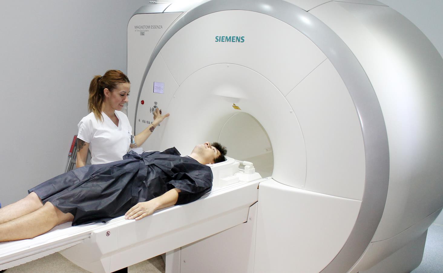
Today, MRI is predominantly used for visualizing soft tissues. It is commonly employed in diagnosing central nervous system (brain and spinal cord) disorders, sports injuries, musculoskeletal conditions such as meniscus tears and herniated discs, as well as in evaluating various neurological diseases.
MRI imaging has not been proven to cause any harm to living organisms. This includes pregnant women; however, it is not recommended to undergo MRI scans during the first trimester when organ development occurs. It is considered unsafe for individuals with metal implants, pacemakers, intraocular metallic foreign bodies, or those who have suffered gunshot wounds (most metals are incompatible with MRI), as it could pose life-threatening risks.
The duration of a magnetic resonance imaging session varies depending on the region being examined, the number of regions, and the preliminary diagnosis, typically ranging between 15 to 75 minutes.
CT (Computed Tomography)
During a CT scan, an X-ray source rotates 360 degrees around the patient while detectors arranged along the gantry capture the portion of the X-ray beam passing through the body. The data collected is then processed by a computer to generate sequential cross-sectional images of the tissues, which can be viewed on a computer screen.
The technician guides the patient to lie on the examination table either on their back or front, depending on the area to be examined. It’s crucial for the patient to remain still throughout the procedure. CT scans may vary based on the patient’s medical condition and the body part being examined. All CT staff are adequately trained and certified to provide the best possible service to patients.
Mammography
Mammography is used to detect tiny abnormalities in the breast that may not be noticeable through physical examination, making it crucial for diagnosing breast cancer. While mammography can be performed on women of all ages if medically necessary, asymptomatic women are advised to undergo mammography at least once between the ages of 35 to 40.
During the procedure, the breast is gently compressed between two layers of the device for a few seconds. Therefore, it’s recommended to schedule mammograms when the breasts are least sensitive, particularly for women with tender breasts.
Bone Densitometry
Bone densitometry measures the amount of calcium and other bone minerals in a specific bone area using X-rays. It is often performed on the spine and hip bones and sometimes includes whole-body scans for both children and adults. X-rays are the oldest and most commonly used method for medical imaging, and the ionizing radiation dose during bone mineral density measurement is minimal.
PHYSICIANS IN THIS FIELD

DR. ERHAN CELENK
DR. BULENT SAHIN

Dr. Gülen Safiye TEMEL
![]()
As the first private healthcare institution in the Thrace Region, we contribute to public health without deviating from our principles of treatment with business ethics, honesty, and compassion...
Telephone:
+90 554 405 62 76
+90 282 673 73 73
Mail:
info@corluvatan.com
Address:
Esentepe Neighborhood Deva Street 11/A Çorlu / Tekirdağ | Turkey